Imaginative and prescient loss among the many aged is a significant healthcare subject: about one in three folks have some vision-reducing illness by the age of 65. Age-related macular degeneration (AMD) is the most typical explanation for blindness within the developed world. In Europe, roughly 25% of those 60 and older have AMD. The ‘dry’ kind is comparatively widespread amongst folks over 65, and often causes solely gentle sight loss. Nonetheless, about 15% of sufferers with dry AMD go on to develop a extra severe type of the illness – exudative AMD, or exAMD – which may end up in fast and everlasting lack of sight. Happily, there are remedies that may sluggish additional imaginative and prescient loss. Though there aren’t any preventative therapies out there at current, these are being explored in scientific trials. The interval earlier than the event of exAMD might due to this fact characterize a important window to focus on for therapeutic improvements: can we predict which sufferers will progress to exAMD, and assist stop sight loss earlier than it even happens?
In our newest work, printed in Nature Medicine, we collaborated with Moorfields Eye Hospital and Google Health to curate a dataset of photographs of eye retinas, practice a synthetic intelligence (AI) system that would predict the event of exAMD, and conduct a research to judge our mannequin in contrast with skilled clinicians. We reveal that our system is ready to carry out in addition to, or higher than, clinicians at predicting whether or not an eye fixed will convert to exAMD within the subsequent 6 months. Lastly, we discover the potential scientific applicability of our system. Our contribution highlights the potential of utilizing AI in preventative research for illnesses reminiscent of exAMD.
The Moorfields Eye Hospital AMD dataset
We used a dataset of anonymised retinal scans from Moorfields sufferers with exAMD in a single eye, and at high-risk of growing exAMD of their different eye. This contains 2,795 sufferers throughout seven completely different Moorfields websites in London, with illustration throughout genders, age ranges, and ethnicities. These sufferers attend the hospital commonly to obtain remedy, present process high-resolution three-dimensional optical coherence tomography (OCT) imaging of each eyes, at every go to. There’s usually a delay between when exAMD has developed and when it’s identified and handled. To handle this, we labored with retinal consultants to evaluation all scans for every eye and specify the scan when exAMD was first evident.
Coaching an early warning system for AMD
Our system consists of two deep convolutional neural networks that take as enter high-dimensional volumetric eye scans, the place every scan consists of 58 million three-dimensional pixels (voxels). In our previous work, now continuing in collaboration with Google Well being, we developed a mannequin able to segmenting these eye scans into 13 anatomical classes. The segmented knowledge was mixed with the uncooked scan and each had been used as inputs to the prediction mannequin, which was skilled to estimate a affected person’s threat of conversion to exAMD of their different eye inside the subsequent six months.
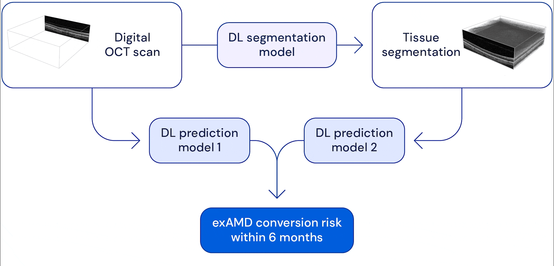
The good thing about a two stage system is that it provides the AI completely different views of the attention scans. Anatomical segmentation of the pictures helps the system study to mannequin dangers based mostly on indicators of identified anatomical indicators reminiscent of drusen (small fatty deposits), or lack of the retinal pigment epithelium (which helps to feed and defend different layers of the retina). Offering the uncooked eye scans permits the mannequin to study to identify different delicate adjustments that would develop into potential threat elements. On the finish, the system combines the knowledge it extracts from these scans to foretell when and if the attention will progress to exAMD inside the subsequent 6 months. We selected this time window to allow the system to foretell at the least two follow-up intervals forward of time, assuming a maximal follow-up interval of three months.
Medical skilled benchmark for future prediction
It’s vital to ascertain a benchmark of skilled human efficiency to match how effectively our system performs to scientific requirements. Nonetheless, prediction of exAMD isn’t a routine job carried out by clinicians, so it’s unclear whether or not this job is even doable. To research this, we performed a research with six retinal consultants – three ophthalmologists and three optometrists, every with at the least ten years of expertise – to foretell whether or not an eye fixed will convert to exAMD inside the ensuing 6 months. Regardless of the duty’s novelty, the consultants carried out higher than likelihood alone – nonetheless, the duty is troublesome, and there was substantial variability between their assessments. Our system carried out in addition to, and in some circumstances higher than, consultants in predicting exAMD development, on the similar time exhibiting much less variability in settlement with every skilled, in comparison with consultants with one another.
AMD is an extremely complicated illness that profoundly impacts the lives of thousands and thousands of individuals all over the world. With this work, we haven’t solved AMD… however I believe we’ve simply added one other massive piece of the puzzle.
Pearse Keane, NIHR Clinician Scientist
Visualising illness development
It might not be sufficient for a system to easily present a prediction: clinicians may additionally ideally search data relating to the anatomic foundation for predictions, which could be of serious use for additional interpretation (for instance, for designing research or contemplating remedies). A good thing about our system is that it routinely segments every scan into identified varieties of tissue. Extracting these anatomical and pathological options gives a scientific methodology to visualise the change in these tissues over time. The danger scores given by our system align with anatomical adjustments over time, and collectively give a richer image of exAMD conversion.

Foresight over hindsight
We’re excited by the potential to assist clinicians and researchers by growing techniques that may assist detect retinal illnesses earlier and inform the scientific understanding of their development. A prediction system reminiscent of this might be used to tell acceptable follow-up intervals to successfully handle high-risk sufferers. Our work builds upon promising early work to develop predictive models for exAMD based mostly on retinal photographs and OCT scans. Since starting our collaboration with Moorfields Eye Hospital in 2016, we’ve printed two promising research highlighting the potential of AI to rework retinal healthcare.
Nonetheless, we all know there’s nonetheless lots to do – this work doesn’t but characterize a product that might be carried out in routine scientific observe. Whereas our mannequin could make higher predictions than scientific consultants, there are various different elements to think about for such techniques to be impactful in a scientific setting. Whereas the mannequin was skilled and evaluated on a inhabitants consultant of the most important eye hospital in Europe, further work can be wanted to judge efficiency within the context of very completely different demographics. A recent study examining the usage of a distinct AI system in a scientific setting highlighted simply a few of the sociotechnical points for such techniques in observe. One other troublesome level to take care of is that any prediction system could have a sure charge of false positives: that’s, when a affected person is discovered to have a situation, or predicted to develop one, that they don’t even have. The tradeoff of including an imprecise AI system to an early warning loop might be unnecessarily expensive to sufferers who aren’t truly in danger, and would should be thought of fastidiously in scientific research of how such techniques could be utilized in observe. On this paper, we suggest two system working factors to steadiness sensitivity (a measure of how effectively it accurately identifies the illness) and specificity (a measure of how low the false constructive charge is). For instance, at a specificity of 90%, a sensitivity of 34% is achieved, that means that the system accurately recognized development in a single third of scans that did go on to progress inside 6 months. This might determine various sufferers at excessive threat with a precision which may be ample to tell research of novel remedy methods which may mitigate imaginative and prescient loss and enhance affected person outcomes.
We want to thank Moorfields Eye Hospital and the clinicians who helped curate the info and had been concerned in our benchmarking research. Please see the paper for all acknowledgements and additional particulars on the work. As well as, we’ve open-sourced the mannequin code for future analysis, out there here, and Moorfields shall be making the dataset out there by way of the Ryan Initiative for Macular Research.